For cationic nanoparticles, the spontaneous nanoparticle−protein corona formation and aggregation in biofluids can trigger unexpected biological reactions. To address this issue, Professor P. Chen’s team at the University of Waterloo presented a biomimetic strategy for camouflaging the cationic peptide/siRNA nanocomplex (P/Si) with single or dual proteins, which exploits the unique properties of endogenous proteins and stabilizes the cationic P/Si complex for safe and targeted delivery. Based on this, human serum albumin (HSA) and apolipoprotein AI (Apo-AI) were selected to construct biomimetic HSA-dressed P/Si (P/Si@HSA) and dual protein (HSA and Apo-AI)-dressed P/Si (P/Si@HSA_Apo), given that the dual-protein camouflage plays complementary roles in efficient delivery. A branched cationic peptide (b-HKR) was tailored for siRNA delivery, and their nanocomplexes, including the cationic P/Si and biomimetic protein-dressed P/Si, were produced by a precise microfluidic technology. The biomimetic anionic protein camouflage greatly enhanced P/Si biostability and biocompatibility, which offers a reliable strategy for overcoming the limitation of applying cationic nanoparticles in biofluids and systemic delivery. This work, entitled “Engineering Biomimetic Protein Camouflage for Delivering Peptide/siRNA Nanocomplexes,” was published in the Journal of the American Chemical Society.
Article Introduction:
We hypothesized that protein corona formation in biofluids is a process of nanoparticle attenuation by proteins in terms of nanoparticle chemical (e.g., surface charges, composition, and functional groups) and physical properties (e.g., surface energy and thermodynamics) to maintain homeostasis. Thus, those protein species with certain functionalities derived from an innate protein corona could be exploited as biomimetic protein camouflages based on high-throughput proteomics. The fundamental rationale is to harness identified protein corona species and design a series of biomimetic protein corona-stabilized nanoparticles, which offers more options and possibilities for robust delivery rather than directly applying the cationic or original nanoparticles in biofluids.
In the preparation of P/Si, the instantaneous binding between b-HKR and siRNA was manipulated by using a microfluidic system equipped with a dual-inlet channel. By varying the microfluidic flow rate from 50 to 2000 μL min−1, the size of the produced P/Si can be tuned in a range of 180 to 90 nm. Although the cationic P/Si in RNase-free water was in nanosize, it rapidly (within 15 min) aggregated into microsize in normal saline, cell growth media, FBS, and particularly in human plasma. The rapid aggregation of cationic nanocomplexes resulting from protein absorption and electrolyte-induced destruction of the electrostatic double layer (zeta potential) has been a common issue of cationic nanoparticles. This unsolved problem prompted in-depth protein corona formation studies of P/Si to understand the underlying biochemical mechanisms so as to address it accordingly in a biomimetic manner (Figure 1). The data reliability of proteins identified by LC−MS/MS was further validated through various techniques, including SDS-PAGE, CD spectra, Nano ITC, etc.
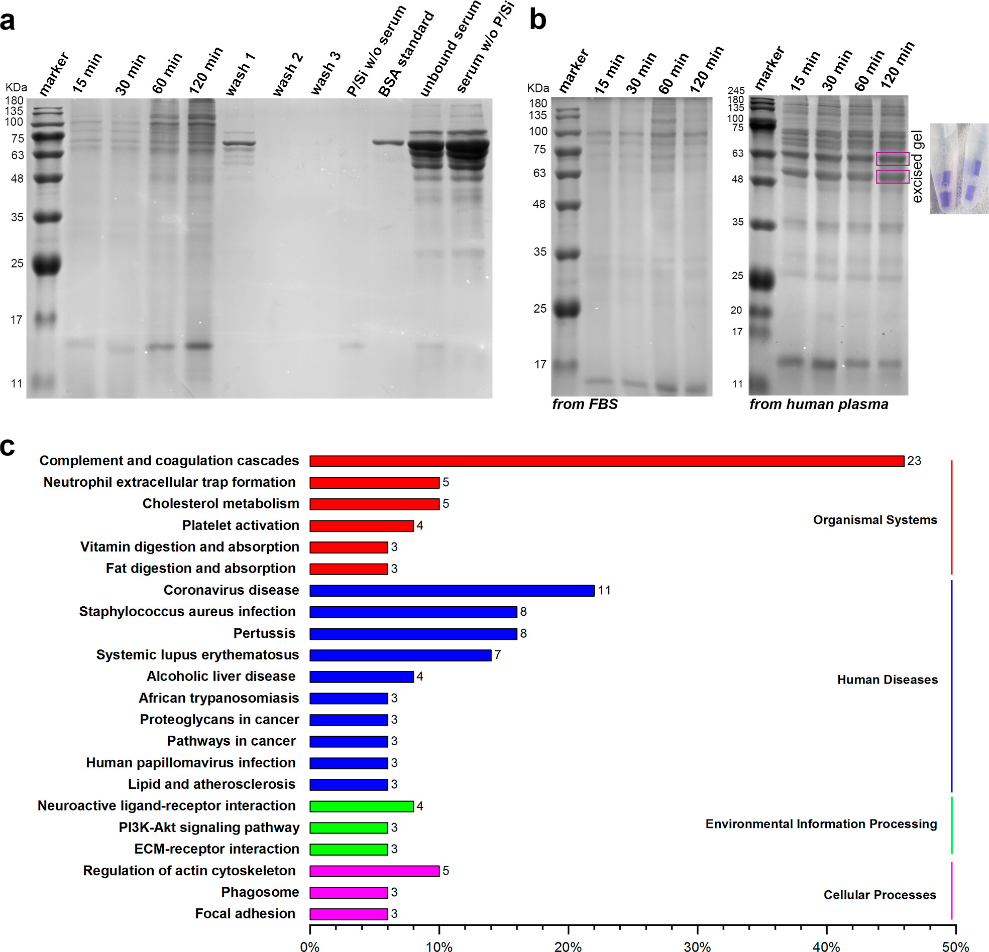
Figure 1. (a) P/Si-PC (FBS) analyzed by SDS-PAGE. (b) Comparison of P/Si-PC separated from human plasma and FBS; these two bands were subsequently excised for further LC−MS/MS analysis. (c) KEGG pathway map of the top 50 identified proteins in P/Si-PC (HP).
These findings indicated that Tf only forms a soft protein corona on P/Si, whereas HSA and Apo-AI can develop into a hard protein corona. The binding affinity of P/Si to HSA was around 10-fold higher than that of P/Si to Tf. According to the multiple-site modeling, it can be estimated that Apo-AI had the highest binding affinity for P/Si among the three proteins. From the above studies, it was speculated that specific domains in HSA can subtly stabilize P/Si. As shown in Figure S3b, the isoelectric point (pI) distribution of the top 50 identified proteins demonstrated that the pI values of the majority of proteins absorbed on P/Si were in the range of 5.5−6.0, which is very close to the pH condition (5.5−6.5) in acidic endosomal vesicles. Therefore, these absorbed proteins are negatively charged under physiological conditions (pH 7.4), and their net charge can theoretically shift to nearly neutral in early endosomes or even positive in more acidic lysosomes (pH 4.5−5.5). These results laid the foundation for leveraging the identified protein corona species to design reversible biomimetic protein camouflages for the cationic P/Si, given the dynamic net charge transformation of these proteins between extracellular conditions and intracellular compartments.
According to these findings and hypotheses, the biomimetic anionic HSA-dressed P/Si (P/Si@HSA) and dual protein (HSA and Apo-AI)-dressed P/Si (P/Si@HSA_Apo) were engineered by microfluidic mixing of cationic P/Si with selected protein corona species. Both P/Si@HSA and P/Si@HSA_Apo were negatively charged under normal physiological pH conditions. The protein-dressed P/Si can maintain their diameters in saline and biofluids but also aggregate immediately under acidic (pH 6) conditions. Since the pI value of Apo-AI is 5.6, the zeta potential of P/Si@HSA_Apo turned to be positively charged when the pH condition was lower than 6.0, which indicated that the protein camouflages were reversible, leading to the exposure of cationic P/Si under acidic conditions, such as in endosomal vesicles. Although the HSA is essential to enhance the biostability of P/Si, the silencing efficiency of P/Si@HSA was compromised compared to that of P/Si, possibly due to its limited endosomal escape. Statistically, there was no significant difference in silencing efficiency between P/Si and P/Si@HSA_Apo for MCF7. These results highlight the advantages and necessity of engineering dual protein-dressed nanocomplexes or more sophisticated reversible protein camouflages to hit a sweet spot, where biostability and biocompatibility can be markedly improved without sacrificing RNAi efficacy.
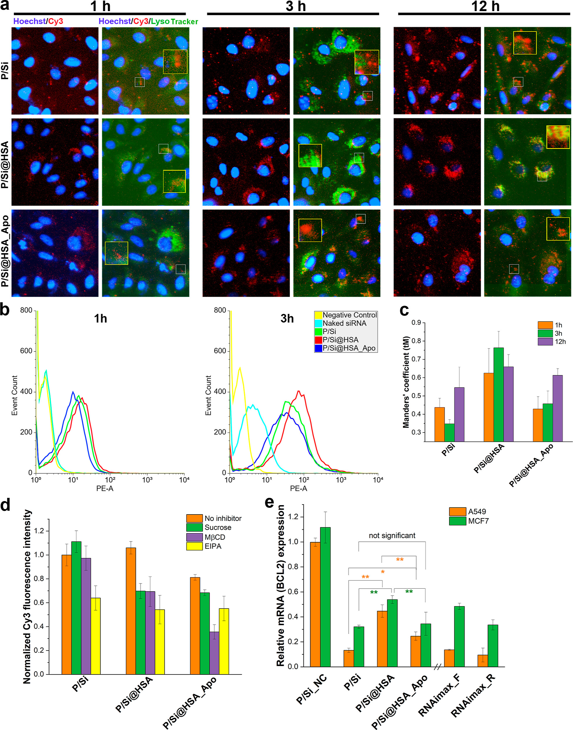
Figure 2. (a) Fluorescence images of cellular uptake and endosomal escape of P/Si and protein-dressed P/Si, with representative areas zoomed in and displayed within rectangles (Cy3-siRNA: red; nuclei: blue; endo/lysosome: green). (b) Cellular (A549) uptake efficiency determined by flow cytometry. (c) Quantification of the Cy3-siRNA colocalizing LysoTracker by calculating the tM values. (d) Cellular uptake with internalization pathway inhibitors. (e) mRNA silencing efficiency of P/Si and nanocomplexes in A549 and MCF7.
The biomimetic protein corona can be engineered as reversible camouflages in physiological conditions according to the nature (pI values, binding affinities, domain structures, etc.) of proteins and physicochemical properties of nanoparticles. This could be a general protocol for engineering protein-stabilized nanoparticles in drug and gene delivery, especially for those with high toxicity and a narrow therapeutic index. The established methodology in this research featured good flexibility, allowing explicit protein selection as camouflage without modification of surface structures or properties of the original nanoparticles.
Article link:
https://doi.org/10.1021/jacs.4c00691